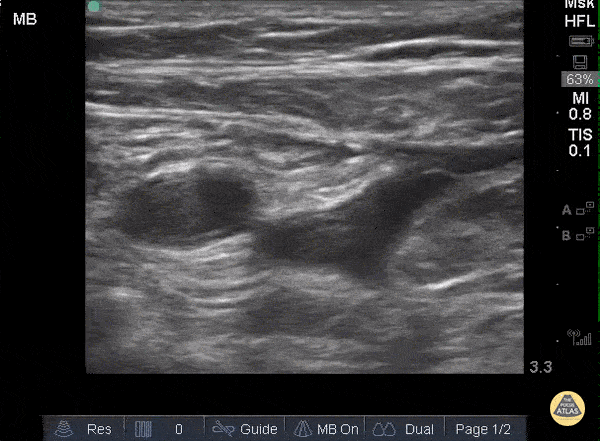
Vascular - DVT - Common Femoral
Caption
When applying pressure to the ultrasound probe, veins collapse first while arteries remain patent and pulsatile. In this clip one can see that proximally, the large pulsatile vessel (which represents the common femoral artery) sits adjacent to a mostly compressible common femoral vein. As the probe moves distally and both the artery and vein split into deep and superficial branches, the superficial femoral vein is not entirely compressible along its medial aspect, representing a thrombus. Sukh Singh, MD