Vascular - VTE in PE
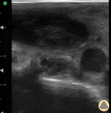
Caption
A 27-year-old women presented to ED with acute onset of dyspnea and chest pain. She was hypoxic with an O2 saturation of 91% on room air and tachycardic. POCUS lung ultrasound revealed an A-line pattern bilaterally and was notably absent for pleural effusions. Venous scan of the proximal right femoral region revealed a non-compressible femoral vein with hypoechoic material within the lumen. High resolution chest CT confirmed diagnosis of PE. Dr. Victor Bang. Emergency Physician at Hospital das Clínicas de Marília. Co-founder of Pocus Jedi. @vmjbang