Vascular - Right Femoral DVT
Caption
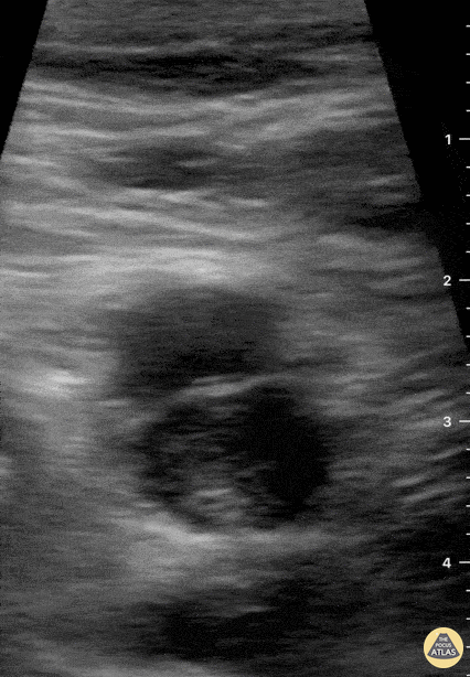
69-year-old male patient with no relevant chronic medical history presents to the ER complaining of two-day right inguinal pain and swollen lower extremity. Directed interrogation revealed one-month subacute dyspnea upon physical effort. Femoral POCUS showed this image. The contractile femoral artery lies superficially and to the left of the screen. The common femoral vein is not fully compressible in this study, and an echogenic thrombus can clearly be identified in its interior. Subsequently, angio CT confirmed a massive bilateral PE, although the patient remained stable and did not require invasive interventions. Dr. Felipe Urriola P. Emergency Unit, Puerto Aysen Hospital. Chilean Patagonia.