Musculoskeletal - Flexor Tendonitis
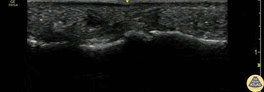
Caption
A 50s M presented with atraumatic finger pain/swelling x2 weeks. On exam, he had focal swelling localized the volar aspect of the ring finger over the proximal phalanx, without fusiform edema/erythema or any limitation of ROM. POCUS showed a small fluid collection adjacent to the flexor tendon. The flexor tendon is shown in long axis, with linear fibers seen just superficial to the bone cortex, and then is seen in short axis. The hypoechoic area superficial to the tendon represents the fluid collection. As the patient had intact ROM and no signs of infection, he was splinted and will follow up for a recheck of tendonitis. Kristy Karkula, PA and Dr. Ruth Foss Denver Health Medical Center