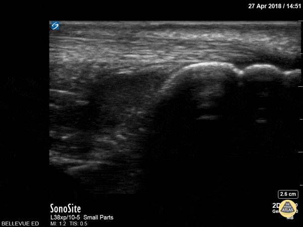
Musculoskeletal - Achilles Tendon
Caption
In this clip we see the linear, fibrillar, echogenic achilles tendon in long axis along the top of the screen. The clip begins distally at the tendon’s insertion onto the calcaneus (the curved hyperechoic structure on the right of the screen). As the probe is moved proximally, the gastrocnemius and soleus muscles become visible in long axis deep to the tendon. Hannah Kopinksi and Dr. Lindsay Davis - NYU Emergency Medicine