OB/Gyn - Tubal Ectopic Pregnancy
Caption
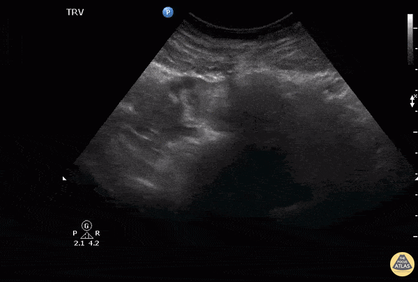
Notice to the left of the screen the presence of an adnexal mass separate to the ovaries which in this case indicates a right tubal ectopic pregnancy. Also noted here within the endometrium is an outer echogenic layer, middle hypoechoic layer and an inner hyperechoic stripe, which makes up the classic trilaminar pattern that is usually observable during the proliferative phase of menstruation. Image courtesy of Robert Jones DO, FACEP @RJonesSonoEM Director, Emergency Ultrasound; MetroHealth Medical Center; Professor, Case Western Reserve Medical School, Cleveland, OH View his original post here