Biliary - Normal Portal Triad - Doppler
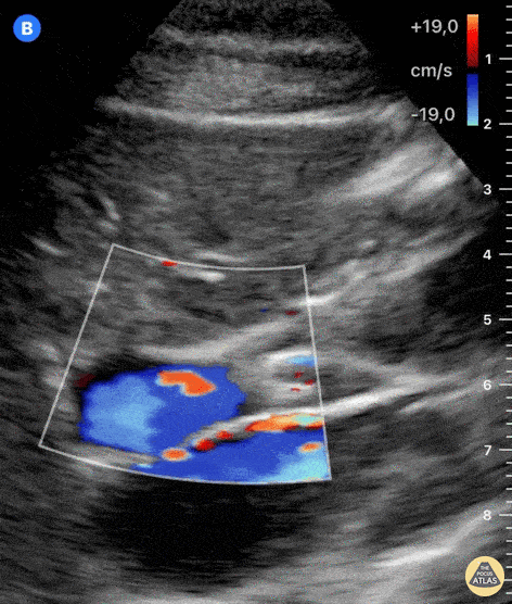
Caption
Both the adjustment of depth and color-doppler allow for better identification of structures. The large, pulsatile inferior vena cava lies at the bottom of the screen. Color-doppler helps to discern blood vessels from other anatomical structures. Anterior to the IVC and from left to right of the screen, we can see the portal vein and hepatic artery with color-flow. Just above the hepatic artery lies another tubular structure which presents hyperechoic walls and an anechoic lumen; this is the common bile duct. Dr. Felipe Urriola P. Emergency Unit, Puerto Aysen Hospital. Chilean Patagonia.