Biliary - Cholelithiasis with Adenomyomatosis
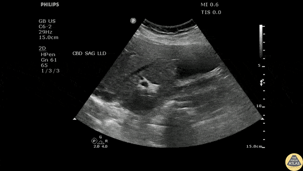
Caption
Longitudinal view of the gallbladder revealing two unique findings. A hyperechoic structure with posterior acoustic shadowing indicative of a stone is located in the neck. Two smaller hyperechoic structures can be seen attached to the wall of the GB body, likely adenomyomatosis. Image courtesy of Robert Jones DO, FACEP @RJonesSonoEM Director, Emergency Ultrasound; MetroHealth Medical Center; Professor, Case Western Reserve Medical School, Cleveland, OH View his original post here