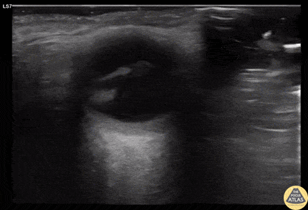
Orbital - Macula Off Retinal Detachment
Caption
70s F with PMH DM, HTN presented to the ED with 1 month of atraumatic, painless vision loss. Her visual acuity was limited to light perception only. POCUS was performed and is shown here. This clip of the eye is obtained with the left of the screen being medial/nasal, and the right of the screen being lateral/temporal. An irregular hyperechoic line is shown in the posterior chamber, which is attached to the optic disk, representing a detached retina. The patient’s presentation with profound painless vision loss, and detached lateral aspect of the retina is consistent with a macula off retinal detachment. Given the timeframe and POCUS findings, the patient was scheduled for urgent but nonemergent ophthalmology follow up for operative repair of the detached retina. Dr. Cody Brevik, PGY4 Denver Health Residency in Emergency Medicine