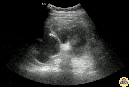
Renal/GU - Severe Hydronephrosis
Caption
30s M PMH HIV, latent TB p/w acute onset flank pain with dysuria. He was found to be febrile and tachycardic. Initial workup was consistent with sepsis due to pyelonephritis. Renal POCUS is shown here, demonstrating severe hydronephrosis, with distortion of the calyceal collecting system as well as thinning of the renal cortex. CT imaging of the abdomen/pelvis demonstrated ureteropelvic junction stenosis causing significant hydronephrosis. Urology was consulted and the patient was admitted for treatment of pyelonephritis as well as further workup of the renal abnormalities. Dr. Nimish Bhatt, US Fellow Denver Health Emergency Ultrasound Fellowship