Pulmonary - Pleural Findings in COVID-19
Caption
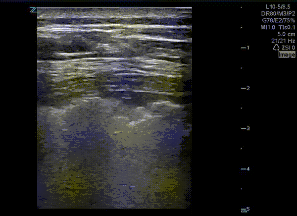
This is a lung ultrasound performed on a patient with COVID-19. The patient had no prior pulmonary disease. They presented with mild tachypnea and hypoxia. A chest x-ray revealed diffuse interstitial and patchy airspace densities. This ultrasound clip shown demonstrates an irregular pleural line with subpleural nodular consolidation and waterfall B-lines. Image courtesy of Dr. Eric Abrams (@eabramsMD)