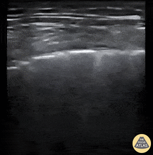
Pulmonary - COVID-19 Pneumonitis
Caption
Patient presented to the Emergency Department with 2-day history of worsening dyspnea and increased work of breathing. He was profoundly hypoxic upon arrival with EMS (O2 sat on 2L via nasal prongs was 42%; improved to 75% upon switching to high flow nasal cannula at 40L/min). A 12-zone lung ultrasound was performed using a linear probe and what is pictured is from L6 (left inferior posterior zone). You can appreciate the coarse irregular pleura, patchy B-lines, and and small areas of consolidation. Findings are typical for clinically-suspected COVID-19 pneumonitis. Cian McDermott, Emergency Physician; Dublin, Ireland @cianmcdermott