Soft Tissue - Peritonsillar Abscess Drainage
Caption
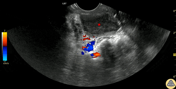
30s F presented with a week of sore throat which had not improved despite corticosteroids and antibiotics. She was referred to the ED by her primary care provider who noted asymmetric swelling in her throat and had concern for peritonsillar abscess (PTA). Evaluation in the ED confirmed this finding, and POCUS was performed, demonstrating a large PTA. The first half of this clip shows a transverse view of the peritonsillar space, where a circumscribed heterogeneous hypoechoic region is seen superficially, which is the PTA (*). Color doppler identifies the carotid artery posterior to the PTA, a critical structure to identify to prevent inadvertent arterial puncture or laceration. The second half of the clip shows the PTA after needle aspiration of 15cc purulence, demonstrating a collapsed abscess cavity with scattered air and significantly reduced abscess size. The patient had immediate relief of her symptoms after needle aspiration, and was discharged with antibiotics and outpatient ENT follow up. Dr. Michael Heffler, PGY-4 Denver Health Residency in Emergency Medicine