Pediatrics - Tracheal Stenosis
Caption
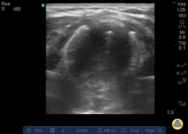
The patient, presenting with stridor, is supine and the airway is seen from the anterior neck in transverse orientation. As the probe is fanned, the bright white line is seen to widen. This column of air moves with inspiration. At its narrowest it is only a few millimeters wide. Growth along the lateral tracheal walls has caused significant tracheal stenosis. In this case, US was used to determine the width of the tracheal column and determine that passage of an ETT would not be feasible. The patient was taken to the OR for an emergent surgical airway. Use of US to estimate tracheal diameter is a novel application. Andrew Liteplo MD, RDMS - Massachusetts General Hospital Chief, Division of Ultrasound in Emergency Medicine Director, Emergency Ultrasound Fellowship