Aorta - Proximal Aorta Anatomy
Caption
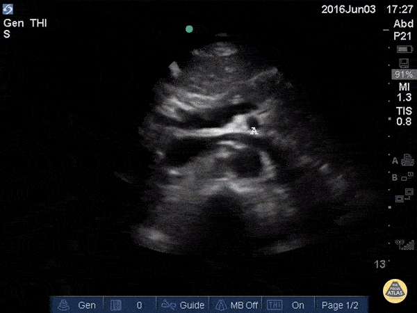
In the beginning of this clip we see several major structures in one view. From superficial to deep: liver, pancreas, splenic vein draining into the portal confluence, superior mesenteric artery in transverse (surrounded by a hyperechoic layer of fat creating the “mantle clock sign”), IVC with the left renal vein branching off, and the abdominal aorta. A vertebral body is visible at the bottom of the screen. As the probe is moved caudally, most of these vessels move out of plane and we are left following the course of the IVC and aorta inferiorly.