IVC & Abnormal Venous Waveforms - Clot At Junction of Right Atrium and IVC
Caption
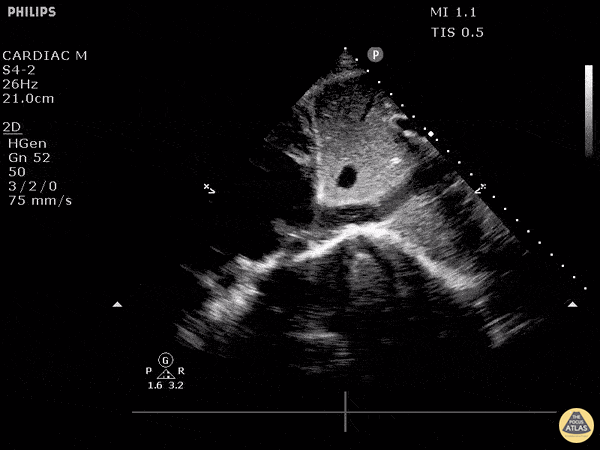
This patient initially presented post-operatively to the emergency department with complaints of dyspnea. As we fan through this saggital view of the IVC as it enters the right atrium, we can see hyperechoic structures suggestive of clot formation. An alternative view of this clot from a subxiphoid view can be seen here. The patient was subsequently diagnosed with a DVT that extended into their central femoral vein, at the same leg that was recently operated on. Image courtesy of Robert Jones DO, FACEP @RJonesSonoEM Director, Emergency Ultrasound; MetroHealth Medical Center; Professor, Case Western Reserve Medical School, Cleveland, OH View his original post here