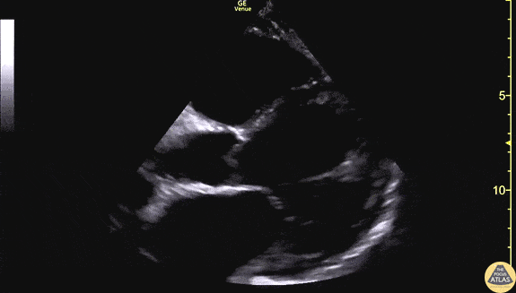
Left Ventricular Dysfunction - Reduced LV Function
Caption
60s M with no known PMH presented to the ED with 3 weeks of progressive dyspnea and bilateral leg edema. POCUS was performed to assess his cardiac function. This clip demonstrates the parasternal long axis and apical 4-chamber views, showing markedly globally reduced LV function, and the endocardium is well seen here. The patient was admitted for further evaluation of his newly diagnosed cardiomyopathy, and formal echocardiography revealed a subtle apical LV thrombus which is not well seen on this POCUS. Dr. Matthew Riscinti Denver Health Medical Center