Right Ventricular Dysfunction - RV Strain with RV dilation and D-sign on PSSA
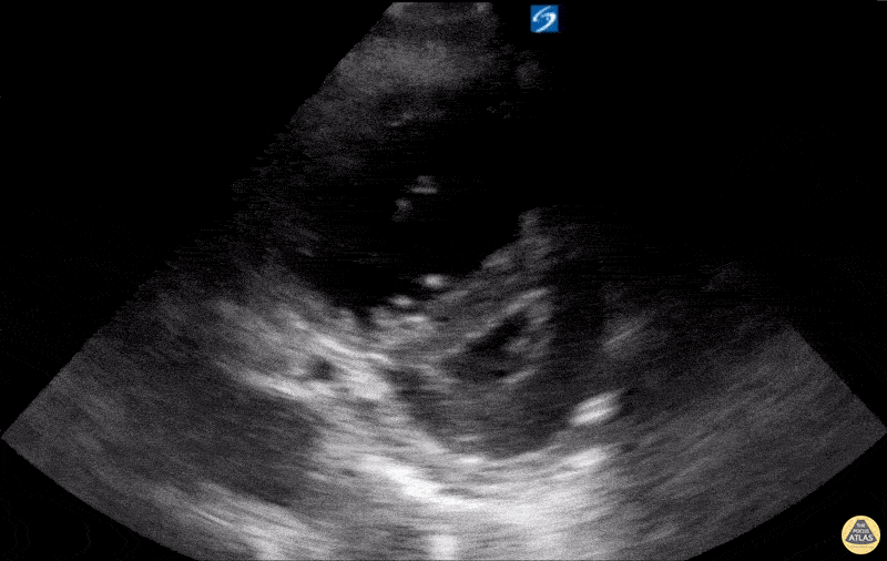
Caption
A 74-year-old male patient with Chronic Obstructive Pulmonary Disease (COPD) (not on home oxygen) presented to the ED with acute worsening dyspnea with persistent hypoxia despite supplemental oxygen, tachypnea, and increased work of breathing. There was no infectious or environmental exposure to explain the patient’s presentation. The patient’s differential diagnosis included acute pulmonary embolism (PE) and COPD exacerbation/progression. Parasternal long-axis (PSLA) and short-axis (PSSA) views showed RV dilatation with RV strain. The RV strain was demonstrated by flattening of the interventricular septum, creating a D-shaped LV during systole (D-sign). Also, it shows hyperdynamic LV from tachycardia with near obliteration of the LV cavity in systole. CT angiography scan showed no evidence of PE. The patient was admitted and diagnosed with a progression of severe COPD with pulmonary hypertension and right ventricular remodeling. Contributed by: Hassan Alshaqaq, MBBS, Emergency Medicine Resident at King Saud University Medical City, @HassanAlshaqaq