Bowel-GI - Appendicitis Longitudinal
Caption
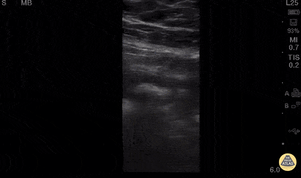
30 y/o M w/ subjective fever, RLQ pain x1day. Tenderness in McBurney’s point w/positive Rovsing’s and Psoas signs, labs significant for mild leukocytosis. POCUS w/ linear probe placed on area of maximal tenderness in RLQ. Transverse and longitudinal views demonstrated non-compressible, blind-ended, non-peristalsing, tubular structure with surrounding hypoechoic free fluid. A central hyperechoic structure representing a fecalith can be appreciated. Outer diameter was measured to be 10mm representing dilated appendix. Findings of acute appendicitis were confirmed with official ultrasound and pt was taken to OR for appendectomy without CT (limiting radiation for young adult). While POCUS is operator dependent and CT is gold-standard for diagnosis of appendicitis in adults, our POCUS findings had higher positive-predictive value given high pre-test probability (classic sx of appendicitis, alternate diagnoses less likely) and optimal pt for study (thin male, no prior surgeries). Dr. Robert Allen and Dr. Matthew Riscinti - Kings County Emergency Medicine