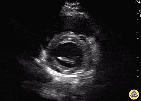
Normal Cardiac Anatomy - Parasternal Short Axis (Mitral Valve) - Normal
Caption
In this view we are evaluating the heart in cross-section. In the center of the screen is the muscular walled left ventricle, which should form a perfect circle. At the beginning of the clip, we see the “fish mouth” appearance of the mitral valve as it opens and closes. Then the probe is tilted inferiorly towards the apex and the papillary muscles come into view. The smaller, thin walled, crescent shaped right ventricle is seen superficially and to the left of the screen. Hannah Kopinski - MS4, Dr. Lindsay Davis - NYU/Bellevue Department of Emergency Ultrasound, Dr. Matthew Riscinti - Kings County Emergency Medicine