Normal Cardiac Anatomy - Parasternal Short Axis - Normal
Caption
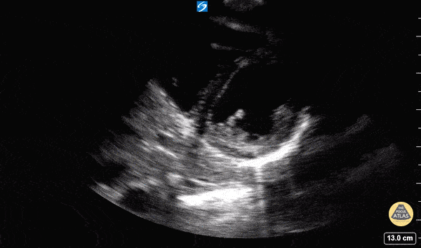
In this view we are evaluating the heart in cross-section. In the center of the screen is the muscular walled left ventricle, which should form a perfect circle. This is at a level below the mitral valve and we can see the papillary muscles come into view. The smaller, thin walled, crescent shaped right ventricle is seen superficially and to the left of the screen. Hannah Kopinski - MS4, Dr. Lindsay Davis - NYU/Bellevue Department of Emergency Ultrasound, Dr. Matthew Riscinti - Kings County Emergency Medicine