Normal Cardiac Anatomy - Parasternal Long Axis - Normal
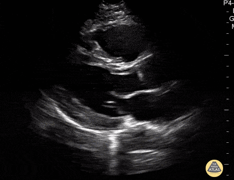
Caption
In this view we see the muscular left ventricle and the smaller left atrium separated by the mitral valve near the bottom of the screen. The aortic outflow tract (valve and root) comes off the left ventricle superficial to the left atrium. The most superficial chamber seen in this view is the right ventricle, separated from the left ventricle by the interventricular septum. The heart is surrounded by the bright hyperechoic pericardium. At the very bottom of the screen, the round structure outside the pericardium is the descending thoracic aorta in transverse view. Hannah Kopinski - MS4, Dr. Lindsay Davis - NYU/Bellevue Department of Emergency Ultrasound, Dr. Matthew Riscinti - Kings County Emergency Medicine