Normal Cardiac Anatomy - Normal Parasternal Long
Caption
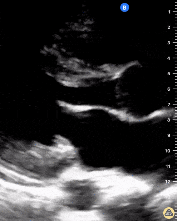
Seen here is a parasternal long axis view of the heart highlighting normal anatomy. You can see the left atrium, aorta, and right ventricular outflow tract, descending thoracic aorta, mitral valve and interventricular septum. Also notice the presence of a small amount of physiologic pericardial fluid. Pericardial fluid in this image is the hypoechoic shadow anterior to the descending thoracic aorta. There is no evidence of pleural effusion, however when present it would appear as a hypoechoic shadow inferior to the descending thoracic aorta. Shahad Al Chalaby, MD. PGY3, Highland Hospital, Alameda Health System Internal Medicine Residency Program @shahad_Chalaby