Normal Cardiac Anatomy - Normal PLAX
Caption
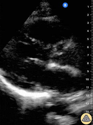
This is a normal parasternal long axis (PLAX) view. The right ventricle (RV) is at the top of the screen. Further down and from left to right: left ventricle (LV), outflow tract, aortic valve, ascending aorta. The actively moving mitral valve separates the LV from the left atrium (LA). At the bottom of the screen, the circular, anechoic image is the descending aorta. Dr. Felipe Urriola. Puerto Aysen Hospital Emergency Department, Chilean Patagonia.