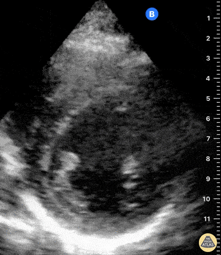
Normal Cardiac Anatomy - Normal Parasternal Short Axis View
Caption
Normal PSAX view at a level of the papillary muscles. In the center of the screen is the muscular walled LV, which forms a perfect circle. The smaller, thin-walled RV is seen superficially and wrapped around the LV. Dr. Felipe Urriola, Puerto Aysen Hospital, Emergency Department, Chilean Patagonia.