Nerve Block Gallery - Erector Spinae Block for Nephrolithiasis
Caption
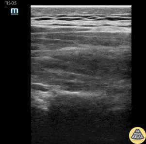
30s M with PMH nephrolithiasis in the past presented with acute onset flank pain. POCUS demonstrated mild hydronephrosis, and his workup was not suggestive of renal insufficiency or UTI, so aggressive symptom control was pursued. He was still experiencing significant pain after two doses of IV morphine, so an erector spinae plane block was offered. After informed consent and setup of supplies, the block was performed and is shown here. The transducer was placed in a sagittal orientation on the mid back at the level of T9, just lateral of midline (toward the affected side) in order to visualize the transverse process. A clip is shown here highlighting the transverse process, and the probe slides laterally to show the more superficial ribs with sliding pleura visible deep to them. The erector spinae muscle can be seen in long axis, just superficial to the transverse process. A 4 inch 20g nerve block needle was advanced using in-plane guidance until the needle tip contacted the transverse process, just deep to the erector spinae muscle. After a negative aspiration for blood, 30 mL of 0.5% bupivacaine with epinephrine plus 1 mL of dexamethasone (4mg in 1 mL) was injected deep to the erector spinae muscle. The patient had improvement of his pain and was able to be discharged with outpatient PCP follow up. Dr. Michael Heffler, Fellow Denver Health Ultrasound Fellowship