Nerve Block Gallery - Posterior Tibial Nerve Block
Caption
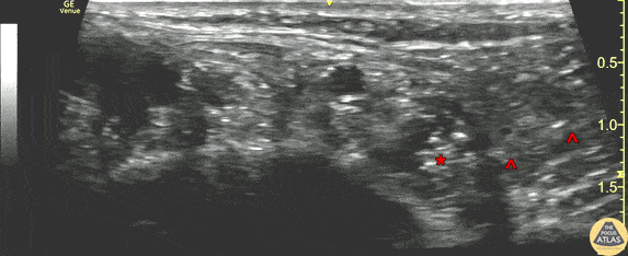
A 20s y/o patient presented after a laceration to the plantar surface of the foot from glass. After exclusion of retained foreign body by XR and physical exam, the laceration was irrigated and repaired using local infiltration of anesthesia, and the patient was discharged. The patient re-presented to the ED within a few hours with continued pain. The laceration repair was intact, and a posterior tibial nerve block was performed for analgesia. This clip shows the nerve block being performed. The linear probe was placed in a transverse plane, just posterior to the medial malleolus, over the “tarsal tunnel.” The left of this image is anterior, and the right is posterior. The needle (^) can be seen entering from the posterior aspect, infiltrating anesthetic around the tibial nerve (*). The pulsating posterior tibial artery is seen anterior to the nerve. The anatomy of the tarsal tunnel can be recalled by the mnemonic “Tom, Dick, And Very Nervous Harry,” which lists the structures in order of anterior to posterior (tibialis posterior tendon, flexor digitorum longus tendon, posterior tibial artery, posterior tibial vein, tibial nerve, flexor hallucis longus tendon), however the tendons and posterior tibial vein are less well seen in this clip. Dr. James Sutton, PGY-3 Denver Health Residency in Emergency Medicine