Nerve Block Gallery - Interscalene Anatomy
Caption
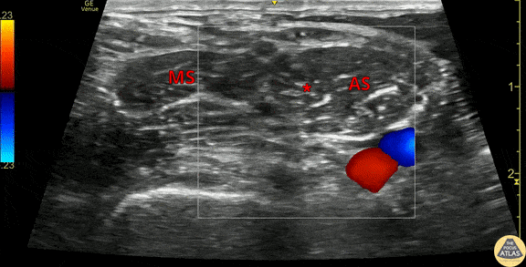
This clip demonstrates the anatomy of the brachial plexus in the interscalene groove. The anterior scalene (AS) and middle scalene (MS) muscles are seen, with the brachial plexus nerve roots (*) seen in the interscalene groove. Color doppler is used to highlight the carotid artery (red) and internal jugular vein (blue) which lie medial/anterior to the brachial plexus. Color doppler is also useful to verify that the brachial plexus nerve roots are not lying deep to other blood vessels. Dr. Michael Heffler, PGY3 Denver Health Residency in Emergency Medicine