Nerve Block Gallery - Interscalene Nerve Root Anatomy
Caption
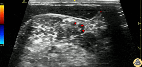
30s M presented with shoulder dislocation. To facilitate reduction, an interscalene nerve block was performed. This clip demonstrates the brachial plexus nerve roots (*) as seen just outside of the interscalene groove - the middle scalene muscle is seen just deep to the nerve roots, with the sternocleidomastoid muscle seen superficial to these. The patient had onset of anesthesia after the block and was able to have closed reduction performed in the ED. Dr. Olivia Serigano, PGY3 Denver Health Residency in Emergency Medicine