Nerve Block Gallery - Fascia Iliaca Nerve Block Anatomy
Caption
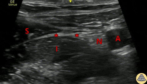
This clip shows the normal anatomy seen during a fascia iliaca nerve block. The image is obtained using a linear transducer in a transverse orientation over the middle to lateral third of the inguinal ligament. The probe marker is to the lateral aspect of the patient. The femoral nerve (N) is seen lateral to the femoral artery (A), and the sartorius muscle (S) is seen superficial to the iliacus muscle (I), with the fascia iliaca in between the two muscles. The target site for injection is just deep to the fascia iliaca, at a point lateral to the femoral nerve (*). Anesthetic should be visualized tracking medially toward the femoral nerve. Dr. Arian Anderson, PGY4 Denver Health Residency in Emergency Medicine