Nerve Block Gallery - Neonatal Fascia Iliaca Block
Caption
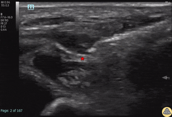
A 50 day-old male was transferred from an outside hospital with concern for NAT after skeletal survey showed old rib fractures, and a significantly displaced and angulated femoral midshaft fracture. A ultrasound-guided fascia iliaca block was performed to aid with passive reduction while in a Pavlik harness. Anesthetic spread can be seen along the fascial plane just superior to the femoral nerve (*). The pulsating femoral artery is seen medial (left of screen) to the nerve. The patient had pain relief with the block, and follow up radiographs showed better reduction after being in the harness without manual manipulation. Dr. Michael Heffler, PGY3, Denver Health Residency in Emergency Medicine Dr. Emily Greenwald, 3rd year PEM fellow, Children’s Hospital Colorado Dr. Megan Mickley, PEM Attending Physician, co-director Pediatric Emergency Ultrasound, Children’s Hospital Colorado