Nerve Block Gallery - Supraclavicular Brachial Plexus Anatomy
Caption
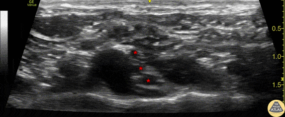
Anatomy of the supraclavicular brachial plexus. This image was obtained with the linear transducer in a parasagittal orientation just lateral to the neck. Left of image is anterior in this view. The brachial plexus (*) is seen just posterior to the pulsating subclavian artery. The hyperechoic first rib can be seen just below the artery and brachial plexus. Drs. Sam Paskin-Flerlage, PGY4 and Michael Heffler, PGY3 Denver Health Residency in Emergency Medicine