Nerve Block Gallery - Superficial Cervical Plexus Anatomy
Caption
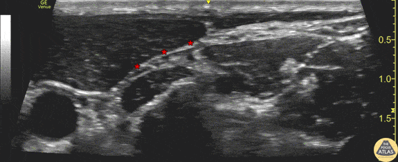
Demonstration of the anatomy of the superficial cervical plexus. The posterior border of the sternocleidomastoid muscle is seen at the center left of screen, and the target fascial plane for a superficial cervical plexus nerve block is highlighted (*). The pulsating carotid artery is seen at the anterior aspect of the image. Drs. Sam Paskin-Flerlage, PGY4 and Michael Heffler, PGY3 Denver Health Residency in Emergency Medicine