Nerve Block Gallery - Serratus Anterior Anatomy
Caption
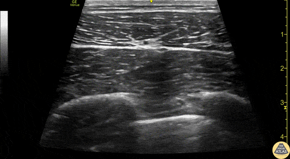
Anatomy of the serratus anterior, as obtained using a linear transducer in a transverse orientation at the level of the 4th/5th rib in the mid axillary line. Anterior is at the left of the image. The latissimus dorsi muscle is seen most superficially, with the serratus anterior muscle deep to it, just above the hyperechoic ribs. Sliding pleura can be seen below the ribs. The thoracodorsal artery can be seen pulsating in the fascial plane between latissimus dorsi and serratus anterior. Drs. Sam Paskin-Flerlage, PGY4 and Michael Heffler, PGY3 Denver Health Residency in Emergency Medicine