Nerve Block Gallery - Tibial Nerve Anatomy
Caption
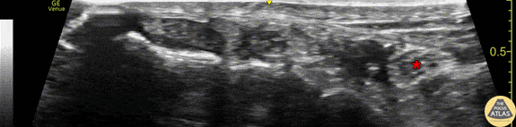
Anatomy of the tibial nerve as it passes posterior of the medial malleolus. The image was obtained using the linear transducer in a transverse orientation just posterior to the medial malleolus. The nerve (*) is seen posterior to (right of) the pulsating posterior tibial artery. Also seen is the hyperechoic malleolar cortex of the distal tibia at the bottom of the image, as well as the tendons of tibialis posterior and flexor digitorum longus anterior to (left of) the artery and nerve. Drs. Sam Paskin-Flerlage, PGY4 and Michael Heffler, PGY3 Denver Health Residency in Emergency Medicine