Nerve Block Gallery - Sciatic Nerve Anatomy
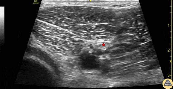
Caption
Anatomy of the sciatic nerve in the popliteal fossa. This image was obtained using the linear transducer in a transverse orientation in the popliteal fossa. The sciatic nerve (*) can be seen superficial to the pulsating popliteal artery, just superficial to the partially compressed popliteal vein. The hyperechoic femoral condylar cortex can be seen at the left of the image. Drs. Sam Paskin-Flerlage, PGY4 and Michael Heffler, PGY3 Denver Health Residency in Emergency Medicine