Nerve Block Gallery - Common Peroneal Nerve Anatomy
Caption
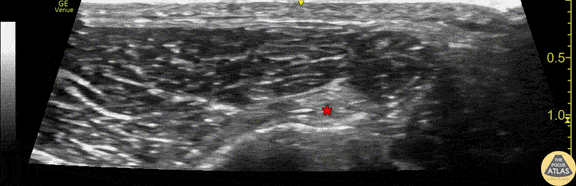
Demonstration of the anatomy of the common peroneal nerve at the level of the proximal fibula. This image was obtained with the linear transducer placed in a transverse orientation on the lateral aspect of the fibular head. The nerve (*) is seen just superficial to the hyperechoic fibular cortex. Drs. Sam Paskin-Flerlage, PGY4 and Michael Heffler, PGY3 Denver Health Residency in Emergency Medicine